

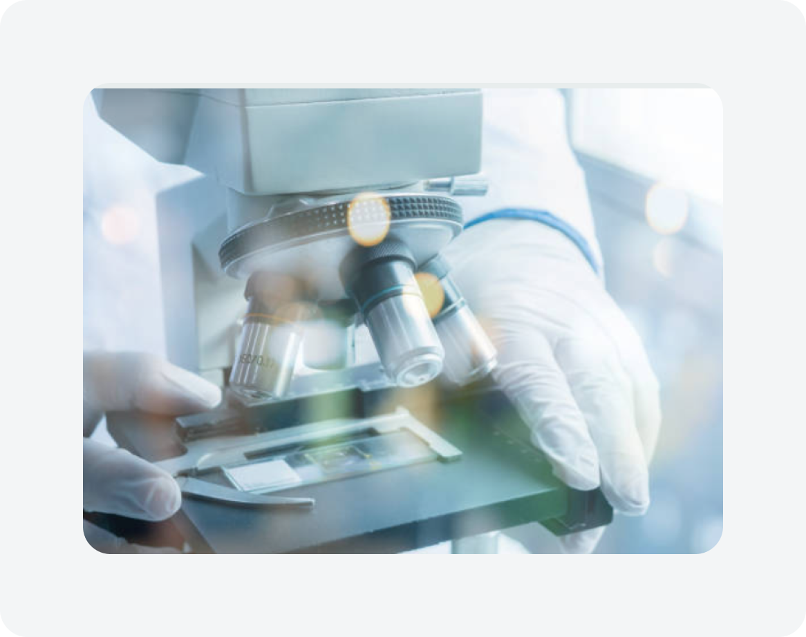
SPOT Imaging’s BlocDoc™ captures macro images of each cut block in as little as two seconds per sample. It also provides the crucial assurance that the entirety of the sample has been documented – providing Pathologists the images they need to verify their slide examinations.
Many reference labs struggle with establishing what they have received from their clients organizations. Most have found that failure to document the incoming samples can lead to their reputation being put into question.
To address this issue may have implemented table top document scanners. These scanners provide a minimum documentation in a workflow that is slow and cumbersome often taking 60 seconds to scan, and save the image files. For large labs this has turned into full time employment for multiple workers producing marginal quality images.
BlocDoc changes the equation by providing a system that is designed for the job automatically capturing and saving enhanced images of labeled cassettes and slides. Placing the cassette or slide takes 2 seconds and the BlocDoc does the rest. The result is a 30x increase in worker productivity freeing worker to perform essential work elsewhere in the lab. The second benefit are professional images that provide tissue detail necessary for conclusive discussions about the samples received bolstering your institution's reputation.
Compact in-hood imaging system supporting in-process documentation
Raw slide tissue levels
Cut-surface of a paraffin block
Below-surface tissue in a paraffin block
Designed to support existing equipment
Foot-pedal camera control eliminates need to remove gloves
Components are sealed, protecting against contaminants
Auto-focus, auto-exposure, auto-calibration camera
Live preview 4K UHD or 720p HD
Captured image 4K UHD or 20 Mp
20x or 8.33x optical zoom
Powered by SPOT Imaging PathSuite Professional software
Supports Telepathology with PathCast software
As a token of our appreciation, we're delighted to offer you a FREE consultation with one of our experienced healthcare professionals. Claim your card now to enjoy these incredible benefits.
%20(1).webp)
.webp)

.webp)
Our system makes it easy for healthcare professionals tho share and collaborate on patient care.

.webp)
